On this page I will present my current and past research by providing a selection of publications.
Optoacoustic mesoscopy
Optoacoustic mesoscopy offers a favorable trade-off between rich optical absorption contrast offered by biological tissues and depth resolution as offered by ultrasound imaging. While most systems were developed for pre-clinical research favoring high resolution offered through optical focusing, I try to push the technology towards usability in clinical settings through acoustic resolution and diffuse illumination. My research in this field focuses mostly on the technical development of fast scanning, multiwavelength imaging systems and advanced reconstruction techniques to recover small features in volumetric images.
- Urs A. T. Hofmann, Weiye Li, Xosé Luís Deán-Ben, Pavel Subochev, Héctor Estrada, Daniel Razansky: Enhancing optoacoustic mesoscopy through calibration-based iterative reconstruction, Photoacoustics 2022: 10.1016/j.pacs.2022.100405
Abstract
Optoacoustic mesoscopy combines rich optical absorption contrast with high spatial resolution at tissue depths beyond reach for microscopic techniques employing focused light excitation. The mesoscopic imaging performance is commonly hindered by the use of inaccurate delay-and-sum reconstruction approaches and idealized modeling assumptions. In principle, image reconstruction performance could be enhanced by simulating the optoacoustic signal generation, propagation, and detection path. However, for most realistic experimental scenarios, the underlying total impulse response (TIR) cannot be accurately modelled. Here we propose to capture the TIR by scanning of a sub-resolution sized absorber. Significant improvement of spatial resolution and depth uniformity is demonstrated over 3 mm range, outperforming delay-and-sum and model-based reconstruction implementations. Reconstruction performance is validated by imaging subcutaneous murine vasculature and human skin in vivo. The proposed experimental calibration and reconstruction paradigm facilitates quantitative inversions while averting complex physics-based simulations. It can readily be applied to other imaging modalities employing TIR-based reconstructions.
- Urs A. T. Hofmann, Sergio Pérez-López, Héctor Estrada, Daniel Razansky: High order pulse-echo (HOPE) ultrasound, Physical Review Applied 2022: 10.1103/PhysRevApplied.17.054024
Abstract
Multiple reflections between transducer and imaged object can naturally occur in ultrasound imaging and other acoustic sensing applications such as sonar. The repeated interaction of the emitted wave front with the imaged object is traditionally regarded as an undesired reverberation artifact, often misinterpreted as fictitious acoustic boundaries. We introduce high-order reflection pulse-echo (HOPE) ultrasound, a method that leverages high-order reflections to improve on several aspects of conventional ultrasound imaging. HOPE is experimentally demonstrated to resolve submicrometer features by breaking through the sampling limit. The major contrast enhancement of the high reflection orders allowed defects within materials invisible to conventional scanning acoustic microscopy to be revealed. The technique is further shown to improve accuracy of frequency-dependent ultrasound attenuation measurements from biological tissues. HOPE ultrasound requires no additional hardware and is easy to implement, underscoring its potential to boost imaging performance in biomedical imaging, nondestructive testing, and other acoustic sensing applications.
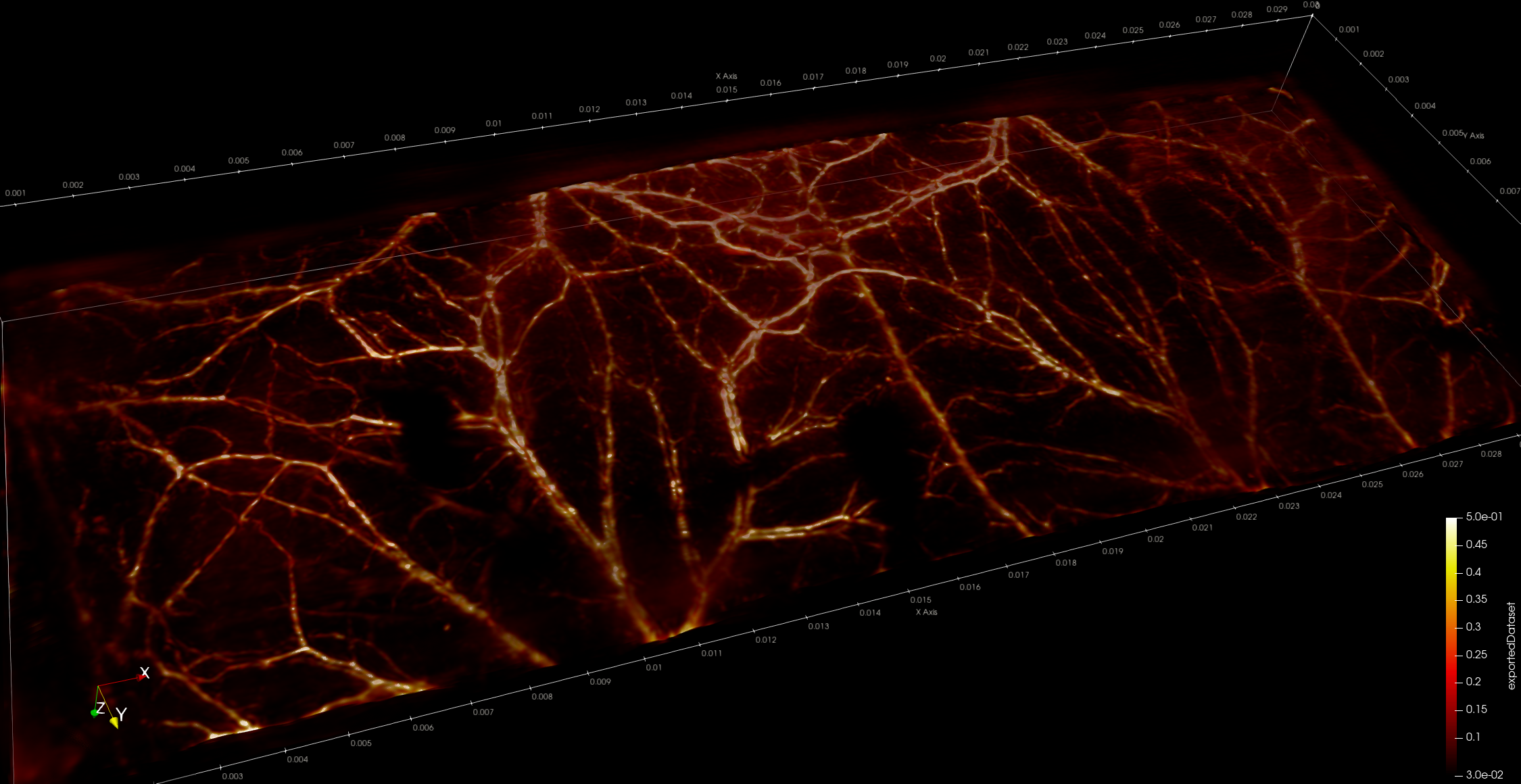
- Weiye Li*, Urs A. T. Hofmann*, Johannes Rebling, Quanyu Zhou, Zhenyue Chen, Ali Ozbek, Yuxiang Gong, Pavel Subochev, Daniel Razansky, Xosé Luís Deán-Ben: Broadband Model-Based Optoacoustic Mesoscopy Enables Deep-Tissue Imaging beyond the Acoustic Diffraction Limit, Laser Photonics Reviews 2022: 10.1002/lpor.202100381
Abstract
Optoacoustic mesoscopy (OAM) retrieves anatomical and functional contrast in vivo at depths not resolvable with optical microscopy. Recent progress on reconstruction algorithms have further advanced its imaging performance to provide high lateral resolution ultimately limited by acoustic diffraction. In this work, a new broadband model-based OAM (MB-OAM) framework efficiently exploiting scanning symmetries for an enhanced performance is presented. By capitalizing on the large detection bandwidth of a spherical polyvinylidene difluoride film while accurately accounting for its spatial impulse response, the new approach significantly outperforms standard OAM implementations in terms of contrast and resolution, as validated by functional in vivo experiments in mice and human volunteers. Furthermore, L1-norm regularization enables resolving structures separated by less than the theoretical diffraction-limited resolution. This unique label-free angiographic performance demonstrates the general applicability of MB-OAM as a super-resolution deep-tissue imaging method capable of breaking through the limits imposed by acoustic diffraction.
- Héctor Estrada, Johannes Rebling, Urs Hofmann, Daniel Razansky: Discerning clavarian microvascular networks by combined optoacoustic ultrasound microscopy, Photoacoustics 19 2020: 10.1016/j.pacs.2020.100178
Abstract
Bone microvasculature plays a paramount role in bone marrow maintenance, development, and hematopoiesis. Studies of calvarian vascular patterns within living mammalian skull with the available intravital microscopy techniques are limited to small scale observations. We developed an optical-resolution optoacoustic microscopy method combined with ultrasound biomicroscopy in order to reveal and discern the intricate networks of calvarian and cerebral vasculature over large fields of view covering majority of the murine calvaria. The vasculature segmentation method is based on an angle-corrected homogeneous model of the rodent skull, generated using simultaneously acquired three-dimensional pulse-echo ultrasound images. The hybrid microscopy design along with the appropriate skull segmentation method enable high throughput studies of a living bone while facilitating correct anatomical interpretation of the vasculature images acquired with optical resolution optoacoustic microscopy.
- Urs A. T. Hofmann, Johannes Rebling, Héctor Estrada, Pavel Subochev, Daniel Razansky: Rapid functional optoacoustic micro-angiography in a burst mode, Optics Letters 45 (9) 2020, 10.1364/OL.387630
Abstract
Optoacoustic microscopy (OAM) can image intrinsic optical absorption contrast at depths of several millimeters where state-of-the-art optical microscopy techniques fail due to intense light scattering in living tissues. Yet, wide adoption of OAM in biology and medicine is hindered by slow image acquisition speed, small field of view (FOV), and/or lack of spectral differentiation capacity of common system implementations. We report on a rapid acquisition functional optoacoustic micro-angiography approach that employs a burst-mode laser triggering scheme to simultaneously acquire multi-wavelength 3D images over an extended FOV covering 50mm×50mm in a single mechanical overfly scan, attaining 28 µm and 14 µm resolution in lateral and axial dimensions, respectively. Owing to an ultrawideband low-noise design featuring a spherically focused polyvinylidene difluoride transducer, we demonstrate imaging of human skin and underlying vasculature at up to 3.8 mm depth when using per-pulse laser energies of only 25 µJ without employing signal averaging. Overall, the developed system greatly enhances performance and usability of OAM for dermatologic and micro-angiographic studies.
- Héctor Estrada, Johannes Rebling, Wolfgang Sievert, Daniela Hladik, Urs Hofmann, Sven Gottschalk, Soile Tapio, Gabriele Multhoff, Daniel Razansky: Intravital optoacoustic ultrasound bio-microscopy reveals radiation-inhibited skull angiogenesis, Bone 133 2020: 10.1016/j.bone.2020.115251
Abstract
Angiogenesis is critical in bone development and growth. Dense, large-scale, and multi-layered vascular networks formed by thin-walled sinusoidal vessels perfuse the plate bones and play an important role in bone repair. Yet, the intricate functional morphology of skull microvasculature remains poorly understood as it is difficult to visualize using existing intravital microscopy techniques. Here we introduced an intravital, fully-transcranial imaging approach based on hybrid optoacoustic and ultrasound bio-microscopy for large-scale observations and quantitative analysis of the vascular morphology, angiogenesis, vessel remodeling, and subsurface roughness in murine skulls. Our approach revealed radiation-inhibited angiogenesis in the skull bone. We also observed previously undocumented sinusoidal vascular networks spanning the entire skullcap, thus opening new vistas for studying the complex interactions between calvarial, pial, and cortical vascular systems.
- Urs A. T. Hofmann, Arne Fabritius, Johannes Rebling, Héctor Estrada, X. Luis Dean-Ben, Oliver Griesbeck, Daniel Razansky: High-throughput platform for Optoacoustic Probing of Genetically Encoded Calcium Ion Indicators, iScience 22 2019: 10.1016/j.isci.2019.11.034
Abstract
Functional optoacoustic (OA) imaging assisted with genetically encoded calcium ion indicators (GECIs) holds promise for imaging large-scale neuronal activity at depths and spatiotemporal resolutions not attainable with existing optical microscopic techniques. However, currently available GECIs optimized for fluorescence (FL) imaging lack sufficient contrast for OA imaging and respond at wavelengths having limited penetration into the mammalian brain. Here we present an imaging platform capable of rapid assessment and cross-validation between OA and FL responses of sensor proteins expressed in Escherichia coli colonies. The screening system features optimized pulsed light excitation combined with ultrasensitive ultrasound detection to mitigate photobleaching while further allowing the dynamic characterization of calcium ion responses with millisecond precision. Targeted probing of up to six individual colonies per second in both calcium-loaded and calcium-unloaded states was possible with the system. The new platform greatly facilitates optimization of absorption-based labels, thus setting the stage for directed evolution of OA GECIs.
- Héctor Estrada, Johannes Rebling, Wolfgang Sievert, Daniela Hladik, Urs A. T. Hofmann, Sven Gottschalk, Soile Tapio, Gabriele Multhoff, Daniel Razansky: Intravital optoacoustic ultrasound bio-microscopy reveals radiation-induced skill vasculopathy, bioRxiv 2019: 10.1101/500017
Abstract
Angiogenesis is critical in bone development and growth. Dense, large-scale, and multi-layered vascular networks formed by thin-walled sinusoidal vessels perfuse the plate bones and play an important role in bone repair. Yet, the intricate functional morphology of skull microvasculature remains poorly understood as it is difficult to visualize using existing intravital microscopy techniques. Here we introduced an intravital fully-transcranial imaging approach based on hybrid optoacoustic and ultrasound bio-microscopy, allowing for large-scale observations and quantitative analysis of the vascular morphology, angiogenesis, vessel remodeling, and subsurface roughness in murine skulls. Our approach also enabled high-throughput physiological studies to understand radiation-inhibited angiogenesis in the skull bone. We observed previously undocumented sinusoidal vascular networks spanning the entire skullcap, thus opening new vistas for studying the complex interactions between calvarian, pial, and cortical vascular systems.
- Quanyu Zhou, Daniil Nozdriukhin, Zhenyue Chen, Lukas Glandorf, Urs A. T. Hofmann, Michael Reiss, Lin Tang, Xosé Luís Deán-Ben, Daniel Razansky: Depth-Resolved Localization Microangiography in the NIR-II Window, Advanced Science 2022: 10.1002/advs.202204782
Abstract
Detailed characterization of microvascular alterations requires high-resolution 3D imaging methods capable of providing both morphological and functional information. Existing optical microscopy tools are routinely used for microangiography, yet offer suboptimal trade-offs between the achievable field of view and spatial resolution with the intense light scattering in biological tissues further limiting the achievable penetration depth. Herein, a new approach for volumetric deep-tissue microangiography based on stereovision combined with super-resolution localization imaging is introduced that overcomes the spatial resolution limits imposed by light diffusion and optical diffraction in wide-field imaging configurations. The method capitalizes on localization and tracking of flowing fluorescent particles in the second near-infrared window (NIR-II, ≈1000–1700 nm), with the third (depth) dimension added by triangulation and stereo-matching of images acquired with two short-wave infrared cameras operating in a dual-view mode. The 3D imaging capability enabled with the proposed method facilitates a detailed visualization of microvascular networks and an accurate blood flow quantification. Experiments performed in tissue-mimicking phantoms demonstrate that high resolution is preserved up to a depth of 4 mm in a turbid medium. Transcranial microangiography of the entire murine cortex and penetrating vessels is further demonstrated at capillary level resolution.
Hand exoskeleton
Stroke patients often suffer from impaired hand movement and require extensive physiotherapy and assistance during activities of a daily living. The rehabilitation engineering laboratory at ETH Zurich is working on a new hand exoskeleton for home use. The focus of my work was on the improvement of actuation technologies and exploring different types of remote actuation systems.
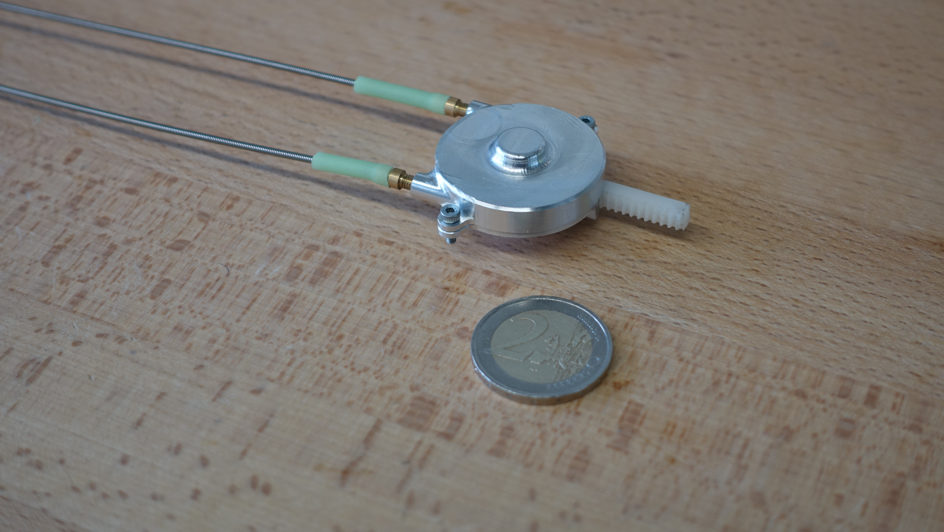
- Jan Dittli*, Urs A. T. Hofmann*, Tobias Buetzer, Olivier Lambercy, Roger Gassert: Remote Actuation Systems for Fully Wearable Assistive Devices: Requirements, Selection, and Optimization for Out-of-the-Lab Application of a Hand Exoskeleton, Frontiers in Robotics and AI, 2021, 10.3389/frobt.2020.596185
Abstract
Wearable robots assist individuals with sensorimotor impairment in daily life, or support industrial workers in physically demanding tasks. In such scenarios, low mass and compact design are crucial factors for device acceptance. Remote actuation systems (RAS) have emerged as a popular approach in wearable robots to reduce perceived weight and increase usability. Different RAS have been presented in the literature to accommodate for a wide range of applications and related design requirements. The push toward use of wearable robotics in out-of-the-lab applications in clinics, home environments, or industry created a shift in requirements for RAS. In this context, high durability, ergonomics, and simple maintenance gain in importance. However, these are only rarely considered and evaluated in research publications, despite being drivers for device abandonment by end-users. In this paper, we summarize existing approaches of RAS for wearable assistive technology in a literature review and compare advantages and disadvantages, focusing on specific evaluation criteria for out-of-the-lab applications to provide guidelines for the selection of RAS. Based on the gained insights, we present the development, optimization, and evaluation of a cable-based RAS for out-of-the-lab applications in a wearable assistive soft hand exoskeleton. The presented RAS features full wearability, high durability, high efficiency, and appealing design while fulfilling ergonomic criteria such as low mass and high wearing comfort. This work aims to support the transfer of RAS for wearable robotics from controlled lab environments to out-of-the-lab applications.
- Urs A. T. Hofmann*, Tobias Buetzer*, Olivier Lambercy, Roger Gassert: Design and Evaluation of a Bowden-Cable-Based Remote Actuation System for Wearable Robotics, IEEE Robotics and Automation Letters 3 (3) 2018, 10.1109/LRA.2018.2809625
Abstract
Wearable robots can assist motor-impaired individuals in activities of daily living, but weight is paramount for usability. Proximally placed actuators and remote actuation systems (RAS) minimize weight on users’ extremities. State-of-the-art RAS employ pneumatics, hydraulics, or Bowden cables, which all have considerable limitations. Here, we present a novel Bowden-cable-based bidirectional RAS featuring high power-to-mass and power-to-volume ratios, easily accessible components, and compact mechanical design. A rack-and-pinion mechanism reduces the force transmitted through the Bowden cables, permitting use of extremely compliant sheaths. The feed-forward friction compensation model, integrated bending angle sensor, and series elastic elements ensure accurate force control across all bending angles of the Bowden cables and the user’s full range of motion. As a proof-of-concept, an RAS was designed for a hand exoskeleton with a maximal output force of 150 N. With a power-to-volume and a power-to-mass ratio of 127 kW/m 3 and 56 W/kg at the output, and of 2.0 kW/m 3 and 1.6 W/kg for the entire system, it outperforms other state-of-the-art RAS. With the implemented speed- and current-limiting, the system operates for at least 2 h continuously. It is water- and dustproof, meeting hygienic and practical demands. Importantly, this novel system can be scaled to the requirements of various applications in wearable robotics.
Modeling the mechanical properties of bone
By modeling the mechanical structures of osteoporotic bone in large scale finite element models, the stability of newly developed implants can be tested efficiently in silico.
- Juri Steiner, Urs A. T. Hofmann, Patrik Christen, Jean M. Favre, Stephen J. Ferguson, G. Harry van Lenthe: Patient-specific in silico models can quantify primary implant stability in eldery human bone, Journal of Orthopaedic Research 36 (3) 2018, pp. 954-962, 10.1002/jor.23721
Abstract
Secure implant fixation is challenging in osteoporotic bone. Due to the high variability in inter- and intra-patient bone quality, ex vivo mechanical testing of implants in bone is very material- and time-consuming. Alternatively, in silico models could substantially reduce costs and speed up the design of novel implants if they had the capability to capture the intricate bone microstructure. Therefore, the aim of this study was to validate a micro-finite element model of a multi-screw fracture fixation system. Eight human cadaveric humerii were scanned using micro-CT and mechanically tested to quantify bone stiffness. Osteotomy and fracture fixation were performed, followed by mechanical testing to quantify displacements at 12 different locations on the instrumented bone. For each experimental case, a micro-finite element model was created. From the micro-finite element analyses of the intact model, the patient-specific bone tissue modulus was determined such that the simulated apparent stiffness matched the measured stiffness of the intact bone. Similarly, the tissue modulus of a small damage region around each screw was determined for the instrumented bone. For validation, all in silico models were rerun using averaged material properties, resulting in an average coefficient of determination of 0.89 ± 0.04 with a slope of 0.93 ± 0.19 and a mean absolute error of 43 ± 10 μm when correlating in silico marker displacements with the ex vivo test. In conclusion, we validated a patient-specific computer model of an entire organ bone-implant system at the tissue-level at high resolution with excellent overall accuracy.